

The gonial angle is the corner where the back of your lower jaw meets its lower border. Clinicians measure it from bone landmarks on dental imaging. A profile photo can show the soft-tissue corner of your jaw, but it cannot provide the same skeletal measurement. If your goal is appearance, compare consistent profile photos and describe the visible contour. Do not seek an X-ray just to check a beauty number.
What the gonial angle measures
The bony landmark at the jaw corner is called the gonion. On a radiograph, an examiner draws one line along the back border of the ascending ramus. A second line follows the lower border of the mandibular body. The angle where those lines meet is the radiographic gonial angle.
The exact construction matters. Radhakrishnan, Sapna Varma and Ajith compared panoramic radiographs with lateral cephalograms in 50 adults in a 2017 Imaging Science in Dentistry study. The two methods produced no statistically detectable difference in their mean angles. Yet a lateral cephalogram superimposes both sides of the jaw, while a panoramic image shows each side separately. Agreement between those two clinical images does not validate a selfie.
| Method | Landmarks or input | What is observable | Main limitation |
|---|---|---|---|
| Radiographic gonial angle | Tangents to the posterior ramus and lower mandibular border | Projected bone outlines | Depends on image type, projection and landmark placement |
| Soft-tissue profile angle | Surface points near the ear, visible jaw corner and chin | Outer profile contour | Skin, muscle and tissue thickness separate it from bone |
| Casual phone photo | Visible pixels or estimated surface landmarks | Appearance in that specific image | Distance, pose, rotation, hair, shadow and lens perspective change the result |
| Front-facing face-shape scan | Frontal 2D outline and landmarks | Visible face outline | It does not show the profile ramus or measure skeletal anatomy |
Why a selfie gives a different answer
Your skin does not mark the gonion like ink on a bone. The masseter muscle sits over the jaw corner. Fat distribution, skin thickness, beard growth and neck position also affect the edge you see. A photo angle is a surface measurement.
Nucera and colleagues tested 96 pretreatment radiographs and 96 profile photographs from people aged 14 to 29. Their 2017 European Journal of Orthodontics study found poor diagnostic concordance between skeletal cephalometrics and both soft-tissue methods. The photo-based and radiograph-based soft-tissue assessments agreed much better with each other. Photography can describe the surface without revealing the underlying skeleton.
Sesqui, Rizal and Suharsini studied 38 children from one defined population. Their 2024 Saudi Dental Journal study found no statistically detectable difference between mean angles from cephalograms and standardized photographs. Those photographs used physical landmark markers, controlled head position, a DSLR camera and same-day imaging. The finding concerns group averages under that protocol. It does not make an unmarked selfie interchangeable with each person's bone measurement.
Close camera distance adds another problem. Derakhshan and colleagues photographed 12 people at several distances and focal lengths. Their 2024 Laryngoscope study found measurable midface distortion at short smartphone distances. They did not test gonial angles, so the study cannot quantify jaw-angle error. It does show why a close selfie is a poor reference for facial proportions.

Illustrative 2D angle calculation
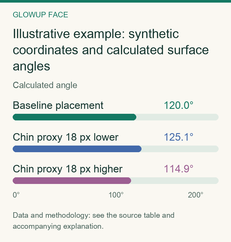
This schematic example shows how three chosen points create a 2D angle. P is an upper-posterior proxy, G is the visible corner, and M is a chin proxy. The calculation uses the angle between vectors P minus G and M minus G. As in standard image coordinates, y increases downward. All coordinates are synthetic and illustrative. They are not measured people, clinical landmarks, product results or expected changes from head movement.
| Scenario | P coordinate | G coordinate | M coordinate | Calculated angle | Change from row 1 |
|---|---|---|---|---|---|
| Baseline placement | (150 px, 213.4 px) | (200 px, 300 px) | (400 px, 300 px) | 120.0° | 0.0° |
| Chin proxy 18 px lower | (150 px, 213.4 px) | (200 px, 300 px) | (400 px, 318 px) | 125.1° | +5.1° |
| Chin proxy 18 px higher | (150 px, 213.4 px) | (200 px, 300 px) | (400 px, 282 px) | 114.9° | -5.1° |
Only the illustrative chin point moves between rows. The synthetic angle shifts by about five degrees. That does not predict what head tilt does to your face. It shows that a 2D result inherits every landmark choice and every projection error.
What high and low mean
Within one fixed method, a high gonial angle is a more open angle. A low gonial angle is a tighter one. Those labels are geometric, not grades. They do not establish attractiveness, health, personality, strength or facial harmony.
There is no evidence-based universal ideal gonial angle. Larrazabal-Moron and Sanchis-Gimeno measured panoramic images from 266 young Mediterranean people for a 2018 Annals of Anatomy study. Values differed across some age and sex groups. The study examined age and sex patterns for forensic use. It did not test attractiveness. Turning a sample average into a beauty target answers a question the study never asked.
The visible jawline adds more variables than the bone angle alone. Two people can have a similar skeletal angle but different surface contours. The reverse can also happen. Lighting and camera position can make one person's corner look sharper in one image and softer in another.

A consistent photo check for appearance
This is an editorial comparison method, not a validated clinical protocol. Its purpose is to make two photos more comparable. It cannot measure bone or diagnose a jaw condition.
- Use the same camera, lens setting, distance and camera height each time. Step back and crop instead of holding the phone close.
- Take a true side profile with a relaxed face. Keep your head level and avoid clenching or pushing your chin forward.
- Use the same plain background and neutral lighting. Keep hair, clothing and facial hair from hiding the jaw corner.
- Compare visible features rather than claiming a bone measurement. Check corner visibility, the ear-to-chin contour and differences between the two sides.
If your real question concerns the visible outline, the on-device face-shape finder estimates the frontal outline. It does not measure the gonial angle or bone. For a change in framing, use the guide to haircuts that soften a jawline. These are appearance decisions, not anatomy tests.
When a clinical measurement is relevant
A dentist or orthodontist may assess the mandibular angle while evaluating growth, bite, asymmetry or treatment planning. That is different from chasing a cosmetic target. Benavides and colleagues' 2026 ADA and AAOMR recommendations say patient history and clinical findings should come before dental imaging. Clinicians should also review existing images and use radiography only when it supports care.
If imaging is already clinically indicated, ask which angle definition and image type the clinician used. Ask whether the value changes a diagnosis or treatment decision. A number that changes nothing is not a reason for extra radiation.
Chewing routines and jaw gadgets should not be sold as ways to set a chosen bone angle. If that is your next question, read what evidence says about chewing gum and face shape. Styling can change the way a contour reads. A selfie, exercise claim or photo scan cannot certify that the mandible itself changed.

Oscar Nascimento
Editorial writer, GlowUp Face
Oscar writes about evidence-based skin care, healthcare, looksmaxxing, and the social and economic research surrounding appearance.
Full profile and all articles →


